Overview
Introduction
Economic Importance
Physical Description
Size
Ecology
Local Occurrence
Global Distribution
Crypsis
Life History & Behaviour
Life Cycle & Reproduction
Locomotion
Feeding (Report)
Anatomy & Physiology
Nervous System
Musculature
Respiration, Circulation & Excretion
Evolution & Systematics
Systematics
Conservation & Threats
Conservation
Threats
References |
LIFE CYCLE & REPRODUCTION
Leptothecate hydrozoans have a biphasic life cycle. This means that they spend part of their life as a hydroid attached to the benthos and the other part of their life as medusae in the water column. The stem of the hydroid colony grows by fixed length budding (Ruppert et al., 2004). It is a polymorphic colony, meaning that it has many zooids responsible for different functions. The gastrozooids are responsible for obtaining nutrients, while the gonangium housing the gonozooid is responsible for producing medusae.
Medusae are released from the gonangium. In the medusa stage, individuals are gonochoristic, meaning that they either male or female. Once fully grown, medusae release eggs or sperm according to their sex. The sperm makes contact with the egg in the water column and a zygote is formed. This zygote develops into a planulae larvae that eventually settles to the benthos and develops into a juvenile hydroid. This cycle then continues.
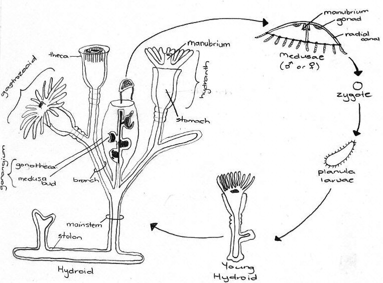
Figure 4.1: Generalised life cycle of a Leptothecatae Hydrozoan. Adapted from
Ruppert,et al., 2004.
|
Species within the genus Aequorea have been observed to reproduce asexually by fission (Ruppert et al., 2004) as shown in Figure 4.2.
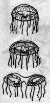
Figure 4.2: Fission in a
hydromedusae. Adapted
from Ruppert et al., 2004 |
In Aequorea sp., the entire lifecycle was not observed. However, observations of the organism under a dissecting microscope showed that it was fully pregnant. This can be seen below:
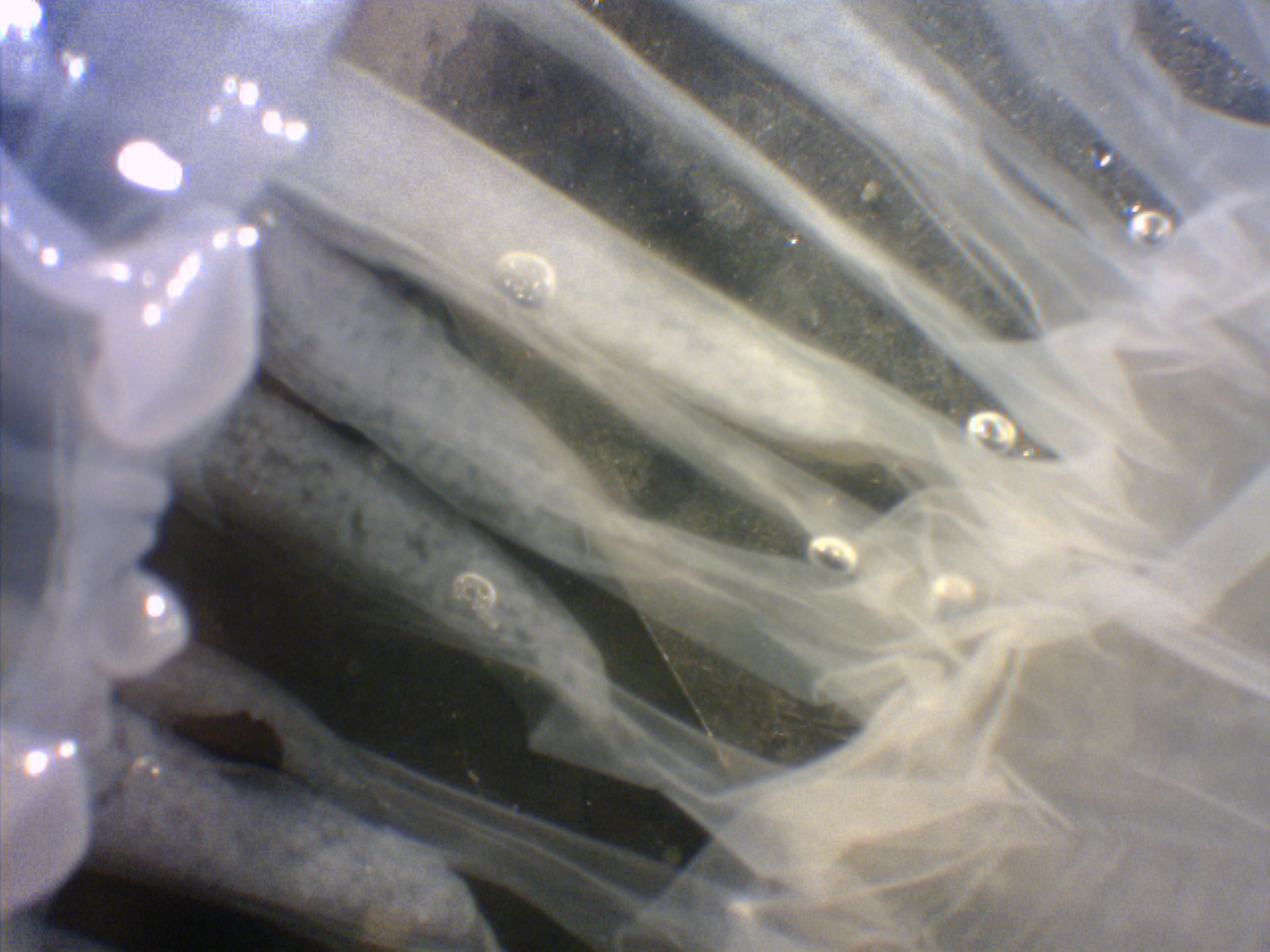
Figure 4.3: Close up of eggs inside Aequorea sp.Photo taken on Heron Island by Katie Maling. |
|
|